CIQTEK to Join Neuroscience 2026 with Volume EM Solutions, USA
October 16, 2026
CIQTEK will join Neuroscience 2026, the annual meeting of the Society for Neuroscience, in Washington, D.C. from November 14 to 18. Visit us at Booth #1014 to explore how high-speed electron microscopy can support large-scale biological imaging, connectomics, and volume electron microscopy workflows.
Neuroscience 2026 will take place at the Walter E. Washington Convention Center and brings together neuroscientists, imaging specialists, technology providers, and researchers from around the world.
High-Speed SEM for Large-Scale Biological Imaging
Modern neuroscience increasingly depends on imaging larger areas, larger volumes, and more complex biological structures.
For volume electron microscopy, imaging speed matters, but speed alone is not enough. Researchers also need stable low-kV performance, reliable image quality, automated acquisition, and workflows that can handle large datasets efficiently.
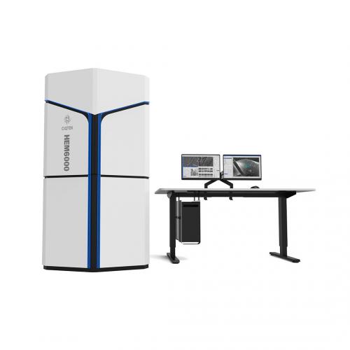
At Neuroscience 2026, CIQTEK will highlight the HEM6000 High-Speed SEM, developed for demanding applications in biological imaging and large-area electron microscopy.
Its capabilities support workflows such as:
High-speed large-area imaging
Low-kV high-resolution observation
Automated image acquisition
Serial section imaging
Volume EM and 3D reconstruction
Neuroscience and connectomics research
The goal is to help researchers collect high-quality structural information faster while reducing the time and effort required for large-scale imaging projects.
Explore CIQTEK Electron Microscopy Solutions
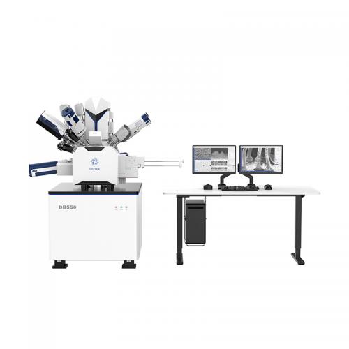
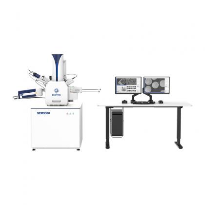
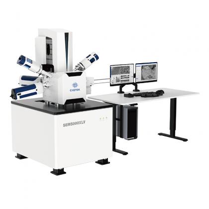
In addition to high-speed SEM, CIQTEK offers a broader electron microscopy portfolio for biological and materials research, including field emission SEM and FIB-SEM systems.
Our team will be available to discuss practical questions around sample imaging, throughput, resolution, automation, and workflow design.
If you are working with brain tissue, neural structures, connectomics, biological ultrastructure, or large-volume imaging, we would be glad to learn more about your research and discuss which electron microscopy approach may fit your needs.
Date: November 14–18, 2026 Location: Walter E. Washington Convention Center, Washington, D.C., USA CIQTEK Booth: #1014
Meet the CIQTEK team at Booth #1014 and see how faster electron microscopy can support your next neuroscience imaging project.
고속 완전 자동화된 필드 방출 주사전자현미경 워크스테이션 CIQTEK HEM6000 고휘도 대구경 전류 전자총, 고속 전자빔 편향 시스템, 고전압 샘플 스테이지 감속, 동적 광학 축, 침지 전자기 및 정전기 콤보 대물 렌즈와 같은 시설 기술을 사용하여 나노 스케일 분해능을 보장하는 동시에 고속 이미지 획득을 달성합니다. 자동화된 작업 프로세스는 더욱 효율적이고 스마트한 대면적 고해상도 이미징 워크플로우와 같은 응용 분야를 위해 설계되었습니다. 이미징 속도는 기존 전계방출 주사전자현미경(FESEM)보다 5배 이상 빠릅니다.
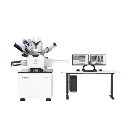
가 + 집속 이온 빔 전계 방출 주사 전자 현미경 그만큼 CIQTEK DB550 집속 이온 빔 주사 전자 현미경(FIB-SEM) 나노 분석 및 시료 준비를 위한 집속 이온 빔 컬럼을 갖추고 있습니다. "슈퍼 터널" 전자 광학 기술, 낮은 수차, 비자성 대물렌즈 설계를 활용하며, "저전압, 고해상도" 기능을 통해 나노스케일 분석 성능을 보장합니다. 이온 컬럼은 Ga를 용이하게 합니다. + 나노 제작 성능을 보장하기 위해 매우 안정적이고 고품질의 이온 빔을 사용하는 액체 금속 이온 소스를 사용합니다. DB550은 나노 조작기, 가스 주입 시스템, 사용자 친화적인 GUI 소프트웨어가 통합된 올인원 나노 분석 및 제작 워크스테이션입니다.